Technology
Digital X Rays

Traditionally, dentists use x-rays to find out what's going on below the surface, developing them in a darkroom full of chemicals, and examining the resulting films on a special light board.
Digital radiography entirely out modes that cumbersome process. Now, a tiny sensor placed in the mouth acts like a miniature VCR camera with an x-ray sensitive chip, exposing you to 50%-90% less radiation exposure than with traditional x-ray techniques. The resulting highly detailed image of your mouth is almost instantaneously translated onto our computer screen, carrying with it all the conveniences of other digitized images. We can rotate it, magnify it, adjust it for contrast, and even color-code it for educational purposes. Because it helps our patients clearly understand the root issues behind their dental health, we're able to work together to determine the very best treatment options for each case.
Hard and Soft Tissue Lasers

Diode laser technology
In dentistry does a clinical tool emerge with so many occasion for treatment as the dental diode laser. These lasers are ideal for procedures involving cutting or from soft tissue, which is why dental diode lasers are also known as soft tissue lasers. Diode lasers are capable of creating precision cuts in gingiva and other soft tissues while also eliminating bleeding at the site and reducing the healing time for the patient. Soft tissue lasers are also ideal for troughing around preparations, sterilizing root canals, treating gum disease and teeth whitening. Many of the latest dental diode lasers are portable and easy to use with simple, touchscreen controls. Take a look and discover how soft tissue dental lasers can enhance every part of your practice.
Intra Oral Camera

This wonderful new technology allows you to relax in your chair while simultaneously observing a real-time pictures of the inside of your mouth magnified beyond normal size on an adjacent computer monitor! Not only does this make it simple to see and understand what the doctor is telling you, but it makes it simple for us to keep incredibly accurate records from one visit to the next.
Cancer Screening

Patients who use tobacco and alcohol heavily are prone to oral cancer. Cancer screening is done in order to detect the signs of cancerous cells at early stage and take necessary steps to cure the same. This can be done during routine dental visit. Dentists make a note of the medical history of patients and check out for abnormal cells using special equipment. The process may involve removal of some of the cells from the mouth and performing biopsy to find out if they are cancerous. The tests performed can help in taking early steps to cure oral cancer.
CBCT

CBCT abbreviated as Cone Beam Computed Tomography is one of the proven technologies for dental medicine. The technology helps in taking 3D digital images of hard bone tissues in teeth and jaws. A cone shaped X-ray beam rotates around the head of the patient to develop a 3D digital image of the dentition. The technology is vastly used in dentistry in recent times because the time taken is much less and the images help in coming up with an effective treatment plan. CBCT can be very helpful when performing dental implant surgeries, orthodontic treatment and other oral surgeries. The complex technology has several advantages and it truly is a boon to the field of dentistry.
Comfort Syringe
Comfort syringe is an electronic device which enables comfortable injection of local anesthetic to patient. The injection is computer controlled. The flow of local anesthesia is regulated which makes the process of injection less painful for the patient. The device has controls to start/stop and increase/decrease the rate at which the local anesthesia is injected. This device is very effective in injecting local anesthesia as compared to manual form of injection.
Velscope
How does the VELscope® work?
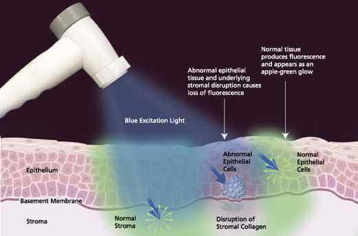
The VELscope® uses Fluorescence Visualization (FV) in an exciting new way. Essentially, bright blue light is shone into the mouth to expose changes and lesions that would otherwise be invisible to the naked eye. One of the biggest difficulties in diagnosing oral cancer is that its symptoms look similar to symptoms of less serious problems. The VELscope® System offers the dentist important insight as to what is happening beneath the surface.
The healthy soft tissue of the mouth naturally absorbs the VELscope® frequency of blue light. Healthy areas beneath the surface of the soft tissue show up green, and the problem areas become much darker.
Here are some of the advantages of using the VELscope® System:
- Can be combined with digital photography.
- Detects lesions, white and red patches.
- Detects problem areas that cannot be seen under white light.
- Exposes precancerous and cancerous tissue.
- FDA-approved.
- Helps dentists check that diseased soft tissue is completely removed.
- Helps diagnose oral cancer in its earliest stages, exponentially increasing the chance of survival.
- Quick, painless examinations.
How is the VELscope® examination performed?
The VELscope® examination literally takes only two or three minutes. It is a painless and noninvasive procedure that saves many lives every single year.
Here is a brief overview of what a VELscope® examination is like:
Initially, the dentist will perform a regular visual examination of the whole lower face. This includes the glands, tongue, cheeks and palate as well as the teeth. Next a pre-rinse solution is swilled around the mouth for slightly less than a minute. The dentist provides special eyewear to protect the integrity of the retinas. The lights in the room are dimmed to allow a clear view of the oral cavity.
The small VELscope® is bent to project blue light inside the mouth. Lesions and other indicators of oral cancer are easily noticeable because they appear much darker under the specialized light.
If symptoms are noted, the dentist may take a biopsy there and then to determine whether or not this is oral cancer. The results of the biopsy dictate the best course of action from there. Otherwise, another oral cancer screening is performed in one year’s time.
If you have any questions or concerns about oral cancer screening or the VELscope® system, please contact our office.
E4D
E4D is a technology we use at our practice which helps us in preparing crowns on the same day. The device uses high-end laser technology to get optical impressions of the prepared tooth. The technology is very accurate and helps to a great extent in designing the crown perfectly for the tooth. A wide variety of other restorations too can be fabricated through the milling unit.


